Terms Used to Describe Ct Results
Standard scores and percentiles describe how a student performed on a test compared to a representative sample of students of the same age from the general population. TOP take-off point.
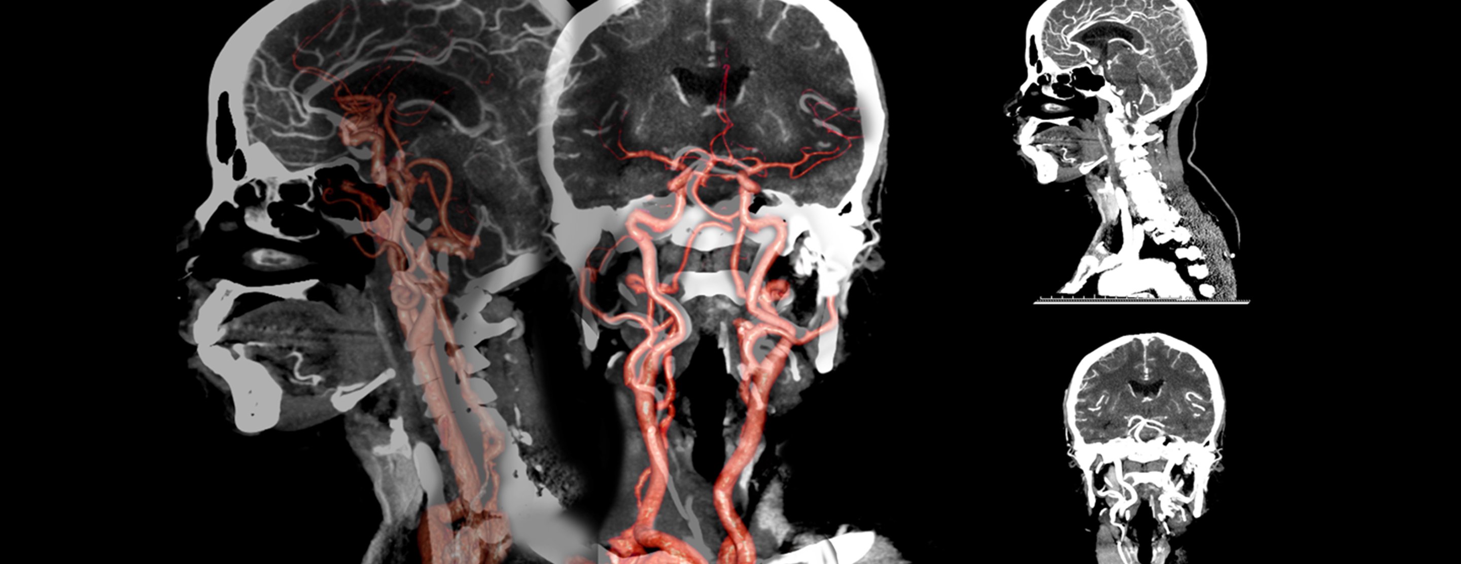
Ct Angiography Head And Neck
Inferior position - lower away from the head.

. Hypoechoic - structures that return weaker echoes of sound waves and appear gray or black on a sonogram. My understanding of a critical result for CTs is a result that indicates action needs to be taken quickly. Infiltrate on a chest X-ray report is a common finding that radiologists use to describe a white abnormal area of unclear cause.
This comparison sample or group is called a norm group. Brighter structures are hyperdense or high density. A pitch greater than 1 will result in an increased dose.
Acute appendicitis ectopic pregnancy new. A radiologist is a doctor who supervises these exams reads and interprets the images and writes a report for your doctor. These values are all the same just with different names.
This refers to subtle thin lines and small dots interspersed throughout the lungs. Language of the Chest X-ray. Inpatie nt Question 2 18 out of 18 points History and exam of the normal newborn infant born in a hospital setting.
There is a tiny calcified granuloma in the right lower lung. Anterior is towards the front of the body Latin. These terms are often found in a radiology report and are used to describe a process in the lungs.
What is the term used to describe the instrumentation and methods used to measure patient dose in CT CT dosimetry TF. Hyperechoic - structures that return greater echoes of sound waves and appear bright white on a sonogram. Refers to acquisition of a volume of CT data.
The term computed tomography or CT refers to a computerized x-ray imaging procedure in which a narrow beam of x-rays is aimed at a patient and quickly rotated around the body producing signals that are processed by the machines computer to generate cross-sectional imagesor slicesof the body. 18 out of 18 points The term used to describe a patient who has been formally admitted to a health care facility is. CT scan images provide more-detailed information than plain X-rays do.
Neighborhoodradiologist September 18 2016 Chest Radiologist Radiology X-ray. A- Image reconstruction algorithms. Darker structures are hypodense or low density.
How to Read Your Radiology Report. That would include readings supporting a diagnosis of. Refer to radiographic findings seen on a CT scan or MRI scan.
A pathologist is a doctor who diagnoses disease byExplaining laboratory testsEvaluating cells tissues and organsThe report gives a diagnosis based on the pathologists examination of a sample of tissue taken from the patients tumor. These test results are important in determining the cause of the pain in addition to the patients specific symptoms and the doctors physical exam results. This report may contain complex words and information.
An 1 cm intraparenchymal hypodensity is also present in the upper pole of the left kidney which does not clearly measure fluid. We can therefore describe things as being high attenuation or low attenuation. It should be kept in mind that all the terms herniated disc pinched nerve bulging disc slipped disc ruptured disc etc.
The patient moves through the gantry with uninterrupted rotation and output of the x-ray tube. C p crossing point. Posterior is towards the back of the body Latin.
Another descriptive term that we commonly use in CT reports refers to the attenuation of tissues which is basically the same thing as density dense tissues will attenuate the xray beam more than low density tissues. The appearance of tissues on a CT scan is described in terms of density. To use the language and results of time series analysis theory to describe from AP 101 at Adventist University of Health Sciences.
Terminology Basic terms of relations. C q quantification cycle. Inpatie nt Selected Answer.
The mathematical techniques used by the computer to reconstruct the computed tomography image are known as. These might be referred to as a CT scan MR scan thyroid scan bone scan etc. Hello my husband recently had an Axial CT the findings are as follows.
A computerized tomography CT scan combines a series of X-ray images taken from different angles around your body and uses computer processing to create cross-sectional images slices of the bones blood vessels and soft tissues inside your body. Radiographic positioning terminology is used routinely to describe the position of the patient for taking various radiographsStandard nomenclature is employed with respect to the anatomic position. Image reconstruction algorithms b.
C t threshold cycle. Answer 1 of 3. A pathology report is a medical document written by a pathologist.
Medical Imaging and Coronavirus COVID-19 Safety. Your doctor sometimes uses medical imaging to diagnose and treat diseases they think you may have. Legend has it doctors add about 10000 new words to their vocabulary in the course of medical training most of which are rarely if ever used outside of medicine.
Term used to describe the computerized images pictures generated by CT MRI ultrasound and nuclear medicine studies. Blood Can Be Very Bad is a mnemonic that can be used when faced with interpreting a CT head scan. Responding to an A2A.
The results of most psychological tests are reported using either standard scores or percentiles. Atelectasis in both of my front tires this week. Before we dive into explaining what a Ct value is we want to take a moment to highlight that that value has been given multiple names over the years including.
In this article we will outline the basic science behind CT scans describe the principles of interpretation and highlight their advantages and drawbacks. An abnormal area of infiltrate on a chest X-ray can represent many abnormalities such as infection water or edema tumor abnormal inflammation not related to infection scarring collapsed lung tissue and other things. This sample of tissue called a specimen is removed during a biopsy.
Computed tomography CT scanning is an extremely common imaging modality in modern medicineWith advancements in technology it is rapidly replacing many diagnostic radiographic procedures. And There is an 14 cm cyst exophytic from the upper pole of the left kidney.
Pin On Aspects评分

Situs Inversus Short Form Of The Latin Situs Inversus Viscerum Is A Term Used To Describe The Radiology Imaging Human Anatomy And Physiology Medical Dental

Leontiasis Ossea A Largely Historical Term Used To Describe A Number Of Conditions Which Result In Radiology Diagnostic Medical Sonography Radiology Imaging


Comments
Post a Comment